Menu
Showing posts with label OTHER PETS. Show all posts


Guinea pigs are by nature smart, cheerful animals, sociable and willing to communicate; both with each other and with their owners. They quickly become trusting, soon listen to their name and enjoy it a lot. The greater the consternation, if one of these animals gets sick. Since we are repeatedly asked by readers, in which concern about a continuous weight loss of the guinea pig is expressed, we have the specialist for small pets of the University of Veterinary Medicine Vienna, Dr. med. Frank Künzel, asked about the topic.

Question: Mr. Künzel, what can be the cause of guinea pigs becoming thinner and thinner?

Dr. Künzel: One possible cause of progressive weight loss in guinea pigs is an overactive thyroid gland. It is typical that the animal decreases, although it eats with great appetite and often shows cravings.

What other symptoms are the pet owner with this disease?

Dr. Künzel: The pet owner noticed changes in guinea pig behavior. Guinea pigs with hyperthyroidism become hyperactive, are nervous and show violent defensive movements when arrested. They take for this species abnormal body postures, sleep, for example, in a lateral position and are difficult to wake. Striking is also that the diseased animal separates from the group, although just guinea pigs are herd animals. Even the shaggy, partly matted hair dress jumps in the eye. But the main reason for the vet visit is in most cases the continuous emaciation of the animal.

How can the vet diagnose "hyperthyroidism"?

Dr. Künzel: The vet first makes a palpation. Since the cause of hyperfunction is often one or more tumors of the thyroid gland, a tumor is felt on the underside of the neck; this can be from the size of a passer-by to a chicken egg. In addition, the veterinarian notices that the animal is breathing too fast, the number of heart beats per minute is too high and the pulse is abnormally strong. Blood must be taken from the guinea pig to determine the amount of thyroid hormone - called total T4. Different methods exist for the determination of this hormone, but it is important that the same method with existing reference levels for guinea pigs is used for the investigations; This is possible, for example, in the central laboratory of the Veterinary University.

Are these tumors on the thyroid gland more benign or malignant?

Dr. Künzel: Studies here at the University have shown that the percentage of benign tumors on the thyroid gland with the malignant scales; About 50% of cases are adenomas and 50% are carcinomas, or cancerous tumors.

Are guinea pigs affected at any age?

Dr. Künzel: No, not at any age, but only animals from the age of three to four years, so middle-aged to older animals.

What treatment options are there?

Dr. Künzel: We have three options: drug therapy, surgery and radioiodine therapy. However, according to the current state of knowledge and the empirical values ​​that we currently have, in most cases drug therapy is to be preferred. The once or twice daily administration of tablets with the active ingredient "Felimazole" inhibits the overproduction of thyroid hormone and affected guinea pigs usually show a rapid improvement. The tablets must be given for life and initially regular veterinary checks must be performed every two to three weeks. Since guinea pigs with hyperthyroidism are often brought to the vet only in advanced disease state, the drug setting difficult and a referral to a special clinic may be necessary. For the surgical removal of the thyroid, there is little experience, the operation involves many risks. The reasons for this are: The risk of anesthesia in the weakened guinea pigs is high and because the thyroid gland is a well-perfused organ, there is also the risk of bleeding. Against the operation also speaks that scattered tumor foci - for example, in the chest - are not detected and therefore it can come to relapse after the surgery has been over. The scintigraphy allows the graphical representation of altered thyroid tissue. For this purpose radioactive pertechnetium is injected intravenously. It accumulates in the tumor and you can see how many "active nodes" are present. The radioiodine therapy takes place immediately after the scintigraphy. The experience with this treatment in guinea pigs is still low, but the results are promising. A disadvantage of this method is that the animals must remain in hospital for up to two weeks after treatment until their radioactivity falls below the legally prescribed limit again. In summary, it can be said that the drug therapy of hyperthyroidism in guinea pigs is well established in practice, as it shows rapid success and is inexpensive and low in risk.
0


Even guinea pigs should be presented to the veterinarian regularly, so that any diseases can be detected and treated at an early stage. Because early detection is the first step to healing.

Dangerous: bridging of the teeth

All guinea-pig teeth, including the molars, are growing throughout their lives. This lifelong growth can be a problem with incorrect feeding. Guinea pigs are naturally used to barren food and require raw fiber-rich feed such as hay and green fodder, which makes intensive grinding movements of the molars necessary. In case of improper diet, e.g. Too much grain, the already strongly tongue-tilted lower molars are rubbed too little, grow across the tongue and eventually form a bridge over the tongue. The affected animals can no longer swallow the food and starve to death at the full bowl. The pet owner is struck by the fact that chewed-up food hangs out of the guinea-pig's mouth and that the animal is heavily salivating and emaciating. For the animal to survive, the teeth must be shortened by the veterinarian.

Most incurable: Guinea pig paralysis

The first symptoms of this dreaded disease are loss of appetite, shaggy coat and severe emaciation. The animals tremble, spasmodic twitching of the back and neck muscles are noticeable. In the further course, the muscles of the hind legs visibly relax and paralysis occurs. Untreated, the animals die after two to ten days, possibly even after three to four weeks. Unfortunately, the treatment is unsuccessful in most cases. Nevertheless, a therapy should be tried in not too weakened animals, since in some cases, but healing can be achieved.

Especially in winter: Lippengrind

Especially in the winter months, many guinea pig owners notice that inflammations with yellow-brown, barky deposits have formed in the corners of the mouth and in the split part of the upper lip. The main reason for this is a lack of vitamin C and vitamin A. Because in contrast to other animals guinea pigs can not produce vitamin C themselves and are therefore dependent on an adequate intake with food. In winter without forage, there is a shortage of vitamin C, as well as vitamin A and unsaturated fatty acids. This leads to a resistance weakness of the skin against bacteria - Lippengrind is the consequence of it. As a therapy, the veterinarian initially delivers high-dose vitamin C as an injection and later as a pet dropper. Local treatments with vitamin A containing ointments and feeding of broken linseed kernels (rich in unsaturated fatty acids) support the healing.

Pododermatitis: inflammation of the bales

Bale inflammations in guinea pigs often indicate deficiencies in attitude. For example, prolonged improper nutrition with grain mixtures instead of raw fiber-rich feeding with hay and green fodder, poorly cleaned stables (urea dissolves the cornea of ​​the paw pads!), Lack of exercise and obesity promote the development of pododermatitis. The paw pads are sore and open, often abscesses. In the therapy, the cancellation of the causes in the foreground. The attitude on a thick hay mat and the treatment of the bales with an antibiotic ointment accelerate the healing.

Cysts on the ovary

The pet owner will notice a circumferential increase in the flank region in combination with symmetrical hair loss. As the cysts can become very large, affected guinea pigs look as if they are highly productive. The ovarian cysts can reach tennis ball size and ultimately displace other organs in the abdomen, which can lead to death. The puncture of the cysts through the abdominal wall brings only short-term relief, as the cysts fill up quickly. With hormonally active cysts and hairlessness on the flanks, a treatment with progestogens can be tried by the veterinarian - the sick animal receives a progestogen injection once a month. Experience has shown that the hair grows quickly after, but the problem of cysts usually persists. The only therapy with certain success is the castration of the guinea pig. This should be done if possible in the initial stage of the disease, as long as the cysts are still small. During castration, both ovaries are removed under general anesthesia.

Hairball in the stomach

The formation of hairballs is promoted by psychological stress on the animals (for example mourning for a companion), boredom and bad grooming by the owner of the animal. Especially during the change of coat regular combing and brushing of the guinea pigs is absolutely necessary, since they usually swallow too much hair when brushing, which then accumulate in the stomach. Also a wrong composition of the food (too little hay and green fodder!) And a too low water intake favor the emergence of hairballs in the stomach. The stomach is thereby expanded, which is why the peristalsis no longer works properly; This means that the food is no longer transported completely from the stomach into the intestine and therefore can not be digested. In smaller hairballs usually conservative therapy with the administration of paraffin oil and subsequent abdominal massages. Large hairballs must be surgically removed.
0


Guinea pigs usually indicate pain by a change in feeding behavior and their cautery habits, the pain often being triggered by a pathological process in the oral cavity. The problem for the attending veterinarian, however, is that the small mouth of a guinea pig is only partially accessible and not easily visible. To study more closely and for treatment, the animal must be anesthetized. Since guinea pigs unfortunately also have an increased risk of narcosis, it is often attempted to correct the teeth when they are awake or to treat superficial dental diseases in a short anesthetic. Very often, however, the cause of toothache lies in the reserve crown (corresponds to the tooth root) and is therefore not visible in the conventional examination of the oral cavity.

Common dental diseases

Incisors and molars are about equally affected by diseases and malformations. But many changes in the area of ​​the incisors have their root cause in the molar area. For example, obliquely worn out incisors often indicate a painful process of the molars. The opening of the first molars of the lower jaw over the tongue is easy to determine when examining the oral cavity. Guinea pigs affected by this can no longer swallow and starve to death without treatment on the full feeding bowl. Other changes in the buccal region that are evident in the exploration of the oral cavity include asymmetric wear, abnormal skewing of the chewing surfaces, tooth tip formation, and dental fractures. In contrast, causal tooth changes, such as macrosontia, a root or a tooth fracture, can only rarely be detected by inspection of the oral cavity. Macrodontia is a tooth that is significantly enlarged in comparison to its physiological form (usually very similar to the tooth of the other side of the body), and represents a mechanical problem for tooth abrasion. The macrodontia is either acquired or already innate. Affected teeth become oversized and are severely inflamed and painful. While problems in the incisor area are already detected during the inspection of the oral cavity, the above-mentioned causative dental diseases can only be diagnosed with imaging techniques.

Clinical study proves necessity of computed tomography (CT)

In a clinical study at the dental clinic of the University of Veterinary Medicine Vienna 66 guinea pigs were examined to what extent computer tomography not only contributes to the safe diagnosis of dental disease, but is unavoidable. The guinea pigs with pain were first subjected to a videoendoscopic examination in the waking state and then subjected to computed tomography. Unlike in the incisor area, where in a bony distension in connection with a tooth fracture, a macrodontia or a tooth loosening in connection with purulent discharge even with the inspection of the oral cavity, the suspected diagnosis "infection of the tooth root" can be made, this is in the molar region not possible - here computed tomography can provide clarity.

Advantages of computed tomography

Since the visible crown area accounts for less than a third of the entire tooth, 85 percent of the painful dental problems occur in the reserve crown area and thus under the gums and can only be detected with a CT examination. This superior examination technique is performed under general anesthesia and is less stressful for the animals compared to the X-ray examination because of the shorter examination time. The conversion into a three-dimensional reconstruction additionally offers an excellent orientation possibility for the operating specialist and also provides information about which animals would benefit from an operation and which not. The result of the clinical study can be summarized in that the sole survey of the oral cavity is not sufficient to diagnose in the guinea pig causally relevant tooth changes such as pathological processes under the gums and the molars, but a computed tomography examination is inevitable to a reliable diagnosis to ask and show the true extent of the disease. Important: The examination by computer tomography is quite affordable: The price for the computed tomography examination of the guinea pig head is currently 120 euros at the university hospital.
0


Examination and therapy same as with dog and cat

Rabbits, guinea pigs, hamsters & co. Were bought mainly as first animal companions for children until a few years ago and unfortunately often treated like toys. Today, however, they have risen to the rank of family members. Even many working adults, for reasons of time, the attitude of a dog or a cat is not possible, have realized how much joy these funny fellow householders. No wonder that there is a desire to enable the small, four-legged companion a long life with a good quality of life. Unfortunately, sometimes the heart breaks even in small pet animals, which severely limits the quality of life. Veterinary help is needed.

Heart built with pets the same

Heart disease in small pets can be diagnosed as well as in dogs and cats, because the heart is basically the same in all these species. However, the examination of these small individuals to the veterinarian even higher demands, because these animals are usually restless and also have an extremely high heart rate, so many heartbeats per minute, have.

Number of heartbeats per minute

Chinchilla: 150 • Guinea pig: 200 • Rabbit: 220
Ferrets: up to 400 • Golden hamsters: up to 412 • Rat: up to 450
Mouse: up to 600

Heart diseases most commonly affect guinea pigs, rabbits, ferrets, chinchillas and African white-bellied iguanas. But also mice, rats and hamsters can be heart disease.

No difference to dog and cat

Small pets are not different from dogs and cats in terms of heart disease. This applies to both the congenital heart disease as well as the acquired.

* For example, in ferrets you often find a congenital heart enlargement, in which the heart muscle dilutes rapidly, which reduces the pumping power of the heart. This weakness of the heart muscle usually only appears from middle age. When the disease is at an advanced stage, pet owners experience symptoms such as shortness of breath, open mouth, apathy, and loss of appetite. In addition, the ferret but also often changes to the heart valves can be found.

* Guinea pigs and rabbits occasionally have a congenital hole in the heart septum. How quickly complaints occur depends on the size of the hole. Most of the affected animals are brought to the veterinarian at the age of one to two years. Fortunately, these defects are usually very small, so they hardly affect life expectancy.

Rabbits and guinea pigs, however, are much more likely to acquire as well as degenerate changes in heart valves between the atrium and the ventricle in the course of life in rabbits and guinea pigs. As with dogs and cats, this leads to the inability of the valves and the backflow of blood either in the body or in the lungs. Here, too, the pet owner notices that the little darling no longer wants to eat, is inactive, hides and breathes quickly with his mouth open.

Examination of the heart

First, the clinical examination of the general condition, followed by the interception of the heart and lungs. The follow-up examinations include ECG and cardiac ultrasound. The same devices are used as for dogs and cats. The x-ray examination of the heart and lungs is often dispensed with, as this represents a risk for the little patients: in a lateral position, the respiratory distress is increased, which can lead to life-threatening conditions. In contrast, the ultrasound examination of the heart can be done in any body position, so is less stressful for the animal.For these studies, the animals do not need to be sedated. The presence of the pet owner can facilitate the investigation if it distracts its little four-legged friend with tidbits. For example, owners of ferrets should have vitamin paste with them; for rabbits it is advisable to bring yoghurt drops or carrots. In some animals, one must also use tricks during the examination: For example, to perform the cardiac ultrasound in the white-bellied urchin, the spiny fellow must be placed in the handstand so that he can not curl up.

Medicines in liquid Form preferred

For the treatment of heart diseases of small pets, the same medicines are used as for dogs and cats: Depending on the clinical picture, digitalis preparations, ACE inhibitors and Lasix. Practical to enter are medications in liquid form.Medicines that are only available in the form of capsules or tablets are almost always too strong for small pets and must be reformulated in the pharmacy into weaker capsules.


0


Hamsters are funny, cute-looking little rodents, but in relation to their height make great demands. According to zoological classification, they belong to the group of the mouse-like and the family of the so-called Wühler. Originally, the small mammals with the big button eyes come from the steppe area of ​​Asia Minor. When in the thirties of the last century the first hamster - who had been caught alive in the context of an expedition to Syria - came to Europe, this caused a real hype in England, whereupon in 1945 a separate hamster club was founded. Since hamsters are dusk- and nocturnal and susceptible to stress, they are not suitable as pets for toddlers.

Generous, well-equipped cage desired

The cage (lattice cage or terrarium) of the small nocturnal mammal needs a quiet location and should be as large as possible despite the small body size and have a minimum of 100x50x50 cm. Hamsters are skilled climbers and live in the wild in an underground branched passage system. A cage with several levels, ladders, tubes, branches of unsprayed fruit trees and numerous hiding places will meet the needs of these rodents. A possibility for sand bathing as well as a situation of bark mulch, branches and dry leaves over the commercial litter round off the offer. The use of hamster wadding should be avoided due to the high accident risk: The small feet can quickly become entangled in the cotton wool and lead from severe injuries to constriction of toes or legs. The legendary wheel must be large enough to prevent spinal damage and be locked on one side.

Special dietary requirements

Hamsters are mainly grain eaters with an increased need for animal protein. Commercially available ready-to-eat food mixes with a low content of high-fat sunflower seeds as well as locust beans, pea flakes and dried herbs correspond to an adequate diet. In addition, once daily fresh food should be offered (for example, some salad, herbs, carrots, corn, peppers and cucumber), once a week, the protein needs by yogurt, pots, mealworms, crickets or dry cat food to be covered, hay and water must always be available.

Different hamster species

In addition to the 100 to 250 g body weight relatively large golden hamster, which occurs in different coat variants (smooth hair, satin, angora, teddy and Rex), there are four particularly popular dwarf breeds: the Chinese striped hamster, the Dsungarian and the Campbellhamster and the Roborowsky dwarf hamsters. With its weight of only 20-30 g, it is the smallest of them all.

Hamsters have anatomical features

On the side of the abdomen, hamsters on both sides have a little hairy circular dark color of the skin, the so-called flank glands, which must not be confused with a plant. They can, however, inflame or degenerate tumorous. The cheek pouches are hamster-type baggy protuberances of the oral mucosa, in which food is stored as a supply in a backpack. Often they are inflamed by wrong posture or nutrition and can not be emptied. The owner then notices a large swelling that can reach from the head to the chest of the hamster, and it is noticeable that the hamster is tired and no longer wants to eat.

Common diseases

Postural disorders include clogged cheek pouches, diarrhea and respiratory distress.

Clogged cheek pouches: The cheek pouches usually have to be emptied and rinsed under anesthesia, the nutrition must be changed over.

Diarrhea: stressed, weakened juveniles that are not kept optimally can develop life-threatening diarrhea, the small stub-tail is humid, and the entire region is heavily polluted with feces; the English term for this is wet-tail disease. Rapid veterinary treatment is needed to relieve symptoms.

Shortness of breath: Favored by poor posture - with too much dust development, lack of hygiene and stress - hamsters can often develop a bacterial bronchitis or pneumonia. The germs involved are streptococci, Bordetella, Pasteurella and mycoplasma. An antibiotic treatment must be done as soon as possible.

Non-postural diseases

Diabetes mellitus: Hamsters, especially the Campbell hamster, have a genetic predisposition to develop diabetes mellitus. High-fat seeds and fruit should therefore not be fed to this species.

Zoonoses (diseases transmissible to humans): Hamsters are usually already infected via the mother with the LCM virus, the causative agent of lymphocytic choriomeningitis. LCM therefore occurs preferentially in younger animals at the age of three to six months. The transfer to humans takes place through direct contact. This virus disease is rare, but can lead to severe, not specifically treatable lung and meningitis. Affected people often have severe flu-like symptoms.

Skin lesions: If a hamster is very restless, does not quite like to eat and constantly scratches - often until the skin is bloody - a mite infestation (such as sarcoptic mange) could be behind it. A precise diagnosis by the veterinarian by microscopic examination of a skin sample (Geschabsel) is recommended to initiate a targeted therapy.

As with us humans, the hamster's teeth are supplied with nerves and blood vessels, and illnesses or injuries to the teeth are painful. But hamster teeth grow lifelong and must therefore be abraded by chewing the food accordingly. In order to support tooth wear and to prevent dental disease, hamsters need raw fiber rich feed, with hay being of particular importance.

As much as hamsters like to stuff their cheek pouches with tasty cereal grains, it can be so bad for the animal's health and teeth. This can lead to the development of back pocket inflammations, which are often aggravated by additional fungal infections and subsequently reduce feed intake. The teeth are therefore no longer sanded off properly and this can cause tooth tips develop very quickly, which then lead to painful injuries of the tongue and cheeks inside. In such cases, the veterinarian must file the tips of the teeth under anesthesia and shorten the teeth to the correct length to allow a complaint-free feed intake. In addition, an accompanying therapy with Antibioticum and Antimycoticum is usually recommended for faster recovery. In some cases, it also causes inflammation of the root tips, which make an extraction of the affected tooth mandatory. Broken teeth also cause severe pain to the animal and can lead to purulent abscesses in the root area and require the extraction of these teeth.
0


Hardly any other part of the organism is discussed as often as the immune system: be it in the many more or less (in) qualified discussions about the pros and cons of vaccinations, or with countless tips for strengthening the immune system. "My PET" spoke to Dr. Michael Leschnik from the University of Veterinary Medicine Vienna on how the immune system works and what its tasks.

Colloquially, the immune system is often referred to as "body police". What is the primary task of the immune system?

Dr. Michael Leschnik: First, the immune system has a monitoring task and, in a figurative sense, an execution task. It should recognize intruders from the outside as well as changes in the body and fight as early as possible. For the most part, one thinks primarily of the fact that the immune system deals with pathogens that invade the organisms from the outside, with foreign proteins of viruses, bacteria or fungi. It is often forgotten that the immune system also plays an essential role in tumorigenesis and prevention. Today, it is known that in the animal as well as in the human being in the course of life tumor cells arise very early on, which are eliminated by the immune system before a cancer arises from it.

What is that, the immune system? We are not dealing with a tangible single organ, but with an absolutely indispensable body function?

Dr. Michael Leschnik: This system, which consists of relatively many interlocking parts, is very complex and undergoes a development that already starts with the puppy, who by no means comes into the world with a fully-fledged immune system. The immune cells are present at birth only to a certain extent and with partly incomplete function, and, moreover, the immune system of a newborn puppy has of course not experienced much. First of all, there is almost no immune memory. That's why the puppy has to get the maternal antibodies in the first one to two days of life with breast milk. This is an important process, because only in the first days of life these antibodies can enter the pancreatic system via the mucous membrane of the gastrointestinal tract. Some of these mother's antibodies work into the twelfth or thirteenth week of life and then slowly break down, while the immune system begins to produce its own antibodies against the pathogens it deals with.

How can you imagine these antibodies?

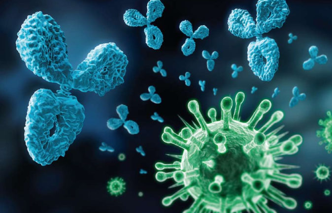
Dr. Michael Leschnik: These are small proteins that usually look Y-shaped. Each antibody forms its own structure with this Ypsilon, which fits to a certain possible intruder. This means that certain antibodies only work against certain pathogens. If such an antibody hits its proper pathogen, it acts like an adhesive. It binds to this pathogen, marking it, so to speak, and this mark eventually becomes the signal for the activation of the further immune system. If the pathogen is eliminated, the immune system stops the production of the corresponding antibody after a short time, because it is currently no longer needed. But it does remember this antibody for a while, and if the organism were then confronted again with the corresponding pathogen, this antibody would be formed much faster and to a great extent. That's the effect we take advantage of in the vaccine. With the vaccination we present to the immune system killed or attenuated pathogens. Then the immune system forms antibodies and at the latest after the 2nd or 3rd vaccination, ie at the end of the basic immunization, a much stronger, faster and more extensive immune response takes place. Depending on the vaccine and disease, these antibodies will last for several months to a few years. This is the basic principle of a vaccine that provides protection against a natural pathogen that could invade the organism.

What happens in the body when it comes to contact with such a pathogen? How does the immune system develop its defenses?

Dr. Michael Leschnik: First of all, it is important to know that the immune reaction in both the fluids, e.g. in the blood, and in the tissues of the body takes place as well as on the body surface. There are immune cells and antibodies that are active in the area of ​​the mucosal surfaces, but they are only the first barrier that sets up the immune system to fend off the intruder. Behind it are the components of the immune system, which spread over the blood stream very quickly throughout the body and can be effective anywhere. Some viruses that we know in dogs and cats are taken up via the mucous membranes. However, this mucosal barrier can be overcome extremely quickly. This first defense on the mucous membrane, especially in the case of viruses, means that they are first recognized by an antibody, labeled and presented to the immune system. Under certain circumstances, however, they can not yet be destroyed. It creates a kind of fight between the different fractions of the immune system and the intruder, which of course does not remain idle. He wants to multiply and not be destroyed, so he trains mechanisms to either escape the immune system, make himself invisible, or look like he's always there and part of the body. Depending on when the immune system can eliminate the pathogen, the disease eventually develops or not. If the immune system can eliminate the pathogen very early, nothing happens that would be visible from the outside, but of course the organism forms the corresponding antibodies. If the organism becomes ill and becomes healthy again, the immune system has won the argument. If this fails, the whole ends either with a permanent illness or, in the worst case, with death.

Does that mean the immune system needs much more than the antibodies for this life-saving battle?

Dr. Michael Leschnik: The first is the antibodies, the second the cells, the so-called cellular defense. In addition, there are many other parts of the chemical-based immune response. For example, the defense cells form proteins that act directly on the pathogen, which can immobilize or destroy it, for example. The two major parts of the immune response, however, always work together. The antibodies are already formed by the immune cells. In addition, there are other cells that primarily have morgues that directly pick up and destroy the pathogens, cells that have labeling properties, or cells that produce substances that harm the pathogens. This is an exciting interaction: Each of the cell types of the immune system has different functions.

Why does the blood count in inflammation result in increased levels of white blood cells, leukocytes?

Dr. Michael Leschnik: The increased levels occur because the immune cells multiply in an inflammation - although we may even have fewer leukocytes at the very beginning of the disease in the blood. It can be imagined as follows: The body is in a peaceful twilight sleep, so to speak. Suddenly something happens, for example in the lungs. First of all, many immune cells migrate from the bloodstream into the lungs. At this time, at the onset of the disease, which is usually accompanied by acute fever, there are even fewer immune cells in the blood. Now, however, the bone marrow, which is responsible for the formation of these immune cells in the body in the adult animal, will produce more of these cells. From the bone marrow, the newly formed cells enter the bloodstream, where we now find many young immune cells. In the course of the disease after a few days, we then find the typical high number of white blood cells.

This entire process of immune defense can also be disturbed. What can lead to the immune system not working optimally?...

Illnesses that require intensive treatment. Allergies are also the result of a misguided immune system. Here suddenly a very massive reaction takes place against a so-called allergen, ie against a substance or part of a substance, which in itself does not have to be harmful, but the misguided immune system decides that this is an intruder and must be fought. Basically, any banal activation of the immune system can trigger such reactions, simple infections, chemical substances, sometimes even medications, and possibly also a vaccine. In the end, the triggers can often not be proven, and of course one can not say that vaccinations are in principle bad, there are so many immune reactions. On the other hand, one demonstrable fact is that the number of serious infectious diseases from vaccinations has decreased significantly.

Strengthening the immune system - that is already a nearly proverbial term today. What does that mean?

Dr. Michael Leschnik: The best way to strengthen the immune system is to always keep it active. A completely sterile environment weakens the immune system, therefore, dealing with the environment and with diseases is not necessarily something negative. Chemical substances that help the immune system in the event of illness, for example, are classic drugs - such as antibiotics - that weaken or kill the pathogen. Vitamins or trace elements support the metabolism necessary for the immune system and can, especially if the system is weakened, have a supporting effect. But a well-functioning immune system, which one must honestly say, we can hardly improve by the administration of any additional preparations. Strengthening the immune system works when it is not quite up to par. Of course, chronic stress, which the animal can not escape, is to be judged negatively. On the other hand, short-term stress is activated and does not have to be detrimental in principle if the whole thing runs in moderation. Also, an infection is stress for the body, but activates and trains the immune system, for example, during vaccination. A trained immune system reacts faster and more effectively, while an overloaded immune system does exactly the opposite. It is not always easy to find the right path. We must avoid fatal and lethal diseases that may be harmful to humans in our animals, but we should only vaccinate against the diseases that really occur and that are relevant to the individual animal. Many vaccinations are done properly for more than a year, so it is not compulsory to vaccinate against every disease every year. The prerequisite, of course, is that the animal does not live in a region where this disease is particularly common. Basically, a single decision has to be made for each patient.

Dr. Leschnik, thank you for the interview!
0


Pug and Bully are affectionate, sensitive, gentle dogs with very good character, who expect a lot of tenderness from their owner. They are pleasant companions for working city dwellers, since they do not need too much exercise, almost never show behavioral problems and do not bite. Since the round, short-skulled head of these dogs corresponds exactly to the so-called Kindchen-Scheme, people who want to see a little bit of "children's set" in the dog are also happy to take a pug or a bully. However, those who choose one of these two breeds should also be aware that both types of breathing are very often respiratory problems that can lead to life-threatening conditions. The "my PET" editorial has been published by Univ. Prof. Dr. Gilles Dupré from the University of Veterinary Medicine, Vienna, asked why mops and bully are so often affected by the serious health problem.

Question: Why is it that the majority of boobs and French bulldogs suffer from massive respiratory problems?

Prof. Dupré: The cause of the problem is the still very popular short-headedness of these races. For by breeding it came to a shortening of the cranial and facial bones, but not to a shortening of the associated soft parts, such as skin and mucous membrane. This means that these animals simply have too much skin and mucous membranes for their short bones, which wrinkles inside and out and obstructs the airways. In addition, there is a malformation of the turbinates, shortened nasal passages and a narrowing of the nostrils in these breeds. Despite animal welfare relevant objections, the breeding program of these breeds was not really changed. The more pugs and French bulldogs are bred, the more problems there are. Here at Small Animal Surgery, we did a study comparing the two breeds in relation to this problem. The study found that pugs have an even narrower nasopharynx than French bullys.

What disease symptoms do these dogs show?

Prof. Dupré: The animals produce snoring breathing sounds, tire quickly when they move, and keep heat and stress very poor. In extreme cases, it leads to fainting and suffocation. Computed tomography (CT) shows that almost no air can flow through the nasal passages. But also problems of the digestive tract, such as vomiting and reflux of gastric contents into the esophagus and thereby inflammation of the esophagus and stomach, are causally related to the problems of the respiratory tract. And that is why it comes to the vomiting and to the reflux of the food, because the animals can breathe only with great effort, so that a negative pressure in the airways and in the abdomen develops and the gastric juices are sucked thereby downright in the esophagus. But one has to say that boobs are less affected by the latter problems than bulldogs. Bullys also often suffer from slower gastric emptying - this is the reason why French bulldogs can either choke up or vomit undigested food three to five hours after feeding.

What is the procedure when such dogs are introduced to small animal surgery?

Prof. Dupré: Our research focuses on finding the narrowest part of the respiratory tract to know why the dog is getting so bad. Only then can we take the appropriate measures. Until ten years ago, it was still believed that an over-long soft palate, which narrows the larynx, was the cause of the breathing difficulties. But today we know that it is not about the narrowing of the larynx by a prolonged soft palate, but about the narrowing of the nasopharynx by the thickened soft palate and the thickened mucous membrane in this area. This leaves no room in the nasopharynx for the passage of air, so that the dogs, who naturally breathe through the nose, only the possibility of breathing with open mouth remains. Although I do not want to say that the extended soft palate was not a problem, the problem has shifted due to the fact that the heads are getting shorter and rounder. The main problem now is the too abundant mucous membrane and too abundant connective tissue. Just for comparison: In a German shepherd, the thickness of the soft palate is only half a centimeter, in pug and bully, however, up to two and a half inches! The soft palate is thus compressed between the tongue base and the skull and finds only space in the posterior nasopharynx, which leads to constriction of the same. The animals must breathe with such great effort that an overpressure arises and closes the larynx, so the soft palate and the voice pocket are pulled into the larynx and the laryngeal bone pressed against each other - this life-threatening condition is called laryngeal collapse. In the course of another study, we found that the narrowest part is at the upper edge of the soft palate - by the way, it is even closer to pugs than to bullies.

What treatments are there and what is the purpose of the treatment?

Prof. Dupré: If an animal with such problems is presented to us at the clinic, we first use computed tomography and endoscopy to evaluate where the upper airways are closest. The goal is then to free the nasal passages and the nasopharynx by surgically widening the nostrils and reducing the thickness of the soft palate to create more space in the nasopharynx. In ninety percent of cases, we achieve a significant improvement in the quality of life of the affected animal. In the pug and bully patients today, a shortening of the soft palate is often no longer sufficient, since the main problem is the thickening. The operation must be as early as possible - always before the onset of laryngeal collapse. Narrowed nostrils should be expanded from the age of six months. Also, the thickened soft palate should be operated on the first, at the latest in the second year of life. The newest method is the "soft palate flap flap", in which the tissue responsible for the thickening is excised and the mucous membrane is wrinkled during sewing. How long is the hospital stay?

Prof. Dupré: Computed tomography, endoscopy and surgery are performed on the same day so as not to have to anesthetize the animal several times. The operated dog has to stay at the clinic overnight, where it is continuously monitored and oxygenated. The next day, the patient can go home and after two weeks, again normally charged.

What Can Newly-Owned Pug Owners Do To Help Their Four-Legged Friends Avoid Serious Breathing Problems In Their Lives?

Prof. Dupré: Pugs and French bulldogs should be presented at the age of six months at the clinic so that it can be medically intervened in good time - even before the larynx collapse. And every dog ​​owner should know that snoring is never a breed trait. Unfortunately, studies show that just pug and bully owners do not want to acknowledge the suffering of their animals. Unfortunately, the shortening of the head and the thickening of the soft palate have rather increased and in pugs, the nasal passages have almost disappeared as a result of an upward rotation of the upper jaw caused by the breeding. In pug and bully breeding, measures should finally be taken with the goal that these races can breathe freely again with a "normal" head
0

Author

authorHello, my name is Jack Sparrow. I'm a 50 year old self-employed Pirate from the Caribbean.
Learn More →


Labels